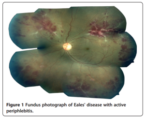
None of the recent studies could differentiate Eales’ disease from tuberculous vasculitis, and also several studies, especially from India where the disease is prevalent, reported the strong association between this type of occlusive vasculitis and tuberculosis.
Thus, we conclude that the name of the disease Eales’ disease will no longer exist.
Category Archives: Resources
Eales disease – current concepts in diagnosis and management
Eales disease – current concepts in diagnosis
and management

A recent article on Eales Disease, received courtesy of Prof. Dr. Jyotirmay Biswas, Director of Uveitis and Ocular Pathology departments – Sankara Nethralaya
PDF article: Eales’ disease -JOII
Abstract
Eales’ disease, first described by the British ophthalmologist Henry Eales in 1880, is characterized by three overlapping stages of venous inflammation (vasculitis), occlusion, and retinal neovascularization. Diagnosis is mostly clinical and requires exclusion of other systemic or ocular conditions that could present with similar retinal features. In recent years, immunological, molecular biological, and biochemical studies have indicated the role of human leukocyte antigen, retinal autoimmunity, Mycobacterium tuberculosis genome, and free radical-mediated damage in the etiopathogenesis of this disease. However, its etiology appears to be multifactorial. The management depends on the stage of the disease and consists of medical treatment with oral corticosteroids in the active inflammatory stage and laser photocoagulation in the advanced retinal ischemia and neovascularization stages.
External link: http://www.joii-journal.com/content/3/1/11
Mycobacterial load in the vitreous of patients
Purpose: To report mycobacterial load in the vitreous of patients labeled as having Eales’ disease.
Methods: Eighty-eight patients were prospectively enrolled into 3 groups: 28 patients with so-called Eales’ disease (group A); 30 positive controls with specific uveitis syndromes (group B), and 30 negative controls (group C). The undiluted vitreous humor samples were collected and subjected to real-time PCR assay for MPB64 gene of Mycobacterium tuberculosis (MTB) and load quantified.
Results: Sixteen (57.14%) vitreous fluid samples in group A; 1 sample in group B, and none of the samples in group C were positive for MTB genome from the vitreous. The copies of MTB genomes in the positive samples in group A were 1.52 × 10⁴ to 1.01 × 10⁶.
Conclusion: MTB genome was demonstrated in more than 50% of vitreous fluid samples with significant bacillary load, indicating that half of patients with so-called Eales’ disease are indeed cases of tubercular vasculitis.
Read More:
Quantitative Polymerase Chain Reaction for Mycobacterium tuberculosis in So-called Eales’ Disease
