
Eales Disease case of a Romanian patient
Case: Romania/2001/M.M./Age18 (Country/Year/Subject/Age)
First noticed symptoms
January 16, 2001 – One evening while I was reading a book, I noticed some points with the left eye (a few, 2,3, close) which were moving with the eye while looking at a page.
First diagnostic – Post uveitis
January 17, 2001 – (next day) I went to the closest hospital (children hospital), at the department of ophthalmology and Dr. Marius TĂTINEANU diagnosed Post uveitis. I was given treatment with two types of eye drops:
- Indocollyre 1 fl.
- Spersalerg 1 fl.
Treatment had no effect.
Next, I went to the Central Military Hospital where I was seen by Dr. Mircea FILIP. and the diagnosis remained the same, but the treatment was changed.
During January to March, the points have appeared also in the right eye, even more than in the left one, then (almost?) completely dissapeared from the left eye and pronounced in the right eye.
Second diagnostic – Both Eyes Retinal Peripheblitis
March 26, 2001 – I went to the ophthalmology hospital where I was saw by Dr. Vasile POTOP. After a few visits (for two or three weeks I was given different generic treatments), the diagnosis was changed from Uveitis to Both Eyes Retinal Peripheblitis, noting that the veins at the back of the eye are “like sausages”. Unable to determine the cause, he also gave me another generic treatment.
Meanwhile points began to disappear from the left eye (don’t know if due to treatment), but were pronounced in the right eye. Then they all disappeared from the left eye.
Treatment:
- Voltaren tb. 25mg x3/day
Reccomended examinations:
- Serological Examination, ???, ???, Toxoplasma.
April 3, 2001 – Cantacuzino Institute, I did the following tests, and the results were good:
- Serological testing for Listeria 1A
- E.L.I.S.A IgG in toxoplasmosis
- Antibodies against Coxiella Burneti, Chlamydia trachtmatis
- Beta hemolytic streptococcus
April 10, 2001 – Control at The Eye Hospital, Dr. POTOP has put the diagnosis of Veins ?xxx?.
Sent to: Neurological Examination, ESR, ?xxx?.
I went to the Children’s Hospital, where I made the following analysis:
- Sinus Radiography
April 11,12, 2001 – I was at the Military Hospital, where I made the following analysis:
- Neurological Exam
- “ASLO”
- Urine
- Field of view
- “VSH”
June 2001 – Visual Field Testing
August 20, 2001 – At the Military Hospital I was seen by Dr. Mihail Zemba, the diagnosis remains Both Eyes Retinal Peripheblitis, and he reccomends it to be seen by Prof. Dr. Benone CÂRSTOCEA.
Treatment:
- Tanakan
- Tarosin
- Diclofenac
Third diagnostic – Retinal Angiomatosis
August 27, 2001 – I was again at the Military Hospital, but I was seen by Dr. Horia Stanca and Prof. Dr. Benone CÂRSTOCEA., who observed that it is more than a Retinal Peripheblitis, and diagnosed Retinal Angiomatosis (ANGIOMATOZA retiniana).
Treatment:
- Sumamed, for 3 days
- Sermion
- Vessel Due
- Voltaren retard, permanently from now on
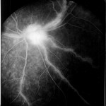
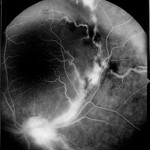
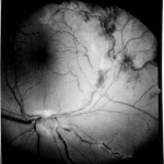
Sent to: LaserOptics Clinic for AFG’s.
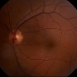
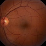
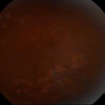
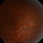
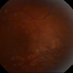
On this same day I went to AFG (angiofluorografie – photographs of the fundus of the eye). The pictures were taken through the process ANGIOFLUOROGRAFIE (I was injected a fluorescent dye that reached the retina in seconds, at which time the pictures were made with a special device).
The scans of the AFG films can be seen below:

First Laser Intervention (Right Eye)
August 30, 2001 – I went through another consultation and I suffered first laser operation: LASER TERMOCOAGULATION in the Right Eye. Before the first surgery I could see many small dots in the right eye and left-bottom side saw unclear.
September 05, 2001 – After the first laser operation, in approx. 4-5 days, something happened in the right eye: a big spot appeared right in the middle of the eye, which put me in the situation that right eye can not read, see something however beside the spot, and right-side-up).
If I remember well, this occured after watching two movies consecutively on a relatively small 21″ CRT TV from a distance of 4-5 meters.
Second Laser Intervention (Right Eye)
September 14, 2001 – I had the second laser surgery: Laser Eye TERMOCOAGULATION Law. My doctor said that a big EDEM formed inside the eye and this is why I can not see so well.
Third Laser Intervention (Both Eyes)
September 28, 2001 – In the morning at 8 o’clock, when I came out to light, I noticed that the left eye recurred several points in the extreme left eye (I could see them if I moved the eye more over to the right). At 3:40 p.m., I suffered a third Laser Operation: LASER TERMOCOAGULATION in Both Eyes because there was a start of “Angiomatoza” in the Left Eye too (I told the doctor that I noticed this morning, and he consulted the left eye again). Before surgery, I was consulted by a nurse who asked me to read letters on the panel. It was found that the right eye is much improved (I saw more than five rows, last time failing to read more than two).
September 28, 2001 – I notice that some dots reappeared in the left eye, in the extreme left vision field, after not having any dots from the beggining of the year
September 29, 2001 – At 11:00 am, something happened and my right eye was covered with several large black spots that covered a large part of vision. After some hours I began seeing unclear with the entire eye (probably blood that flowed inside spreaded). At 9:03 p.m. the large black spot decreased in intensity and I saw that there are actually two spots: one in the middle looking like a broken vein (I guess now that is what is called FLOATERS), remained fixed in one end and the other end moves in the direction you move the eye (respecting inertia, and curls, as vertical, left or right). The other spot (in the right part of the eye) can’t figure it out what is probably blood or another vein. However with the right eye I wasn’t able to see much, barely distinguishing objects.
First Cryosurgery (Right eye)
October 2, 2001 – At The Military Hospital, Miss Dr. Monica ARMEGIOIU did me a cryo surgery to the Right Eye.
November 2, 2001 – Vitrectomy for the right eye is recommended
The final intervention on the right eye – Vitrectomy
November 5, 2001 – I was hospitalized in order to support vitrectomy on the right eye, performed by Dr. Benone CÂRSTOCEA and assisted by Dr. Horia STANCA.
First hours of recovery after vitrectomy
The first hours after vitrectomy are supposed to be spent with the head facing down and as patient as possible (because of a inserted silicone bubble that is supposed to press against the retina to help not detaching after intervention) , but for me they were horror. I had a cold, i could not breath well, and the hospital bed I was supposed to sleep in was not tailored for sleeping with the face down. They should had special beds for this recovery, that have a hole cut to put your face onto. So this moments were agitated and I can’t guarantee that I kept my face down absolutely all the time and I don’t know if this and the blood pressure from the agitation contributed to what I was to find next.
First consultation after vitrectomy
When the eye bandage was taken off, I could not see anything, so it was far worse than before vitrectomy. I could not say I had a shock because I did not knew how it should be in the first day and I was told that it takes some time.
In the next days I was told that the retina detached and it looks like “atomic mushroom cloud”. That was it. I was also told that nothing could be done until at least three months passes, as the eye can not suffer another vitrectomy surgery without high risk of athropy.
Leaving hospital after vitrectomy
November 14, 2001 – I left the hospital after a Vitrectomy surgery.
Treatment to be followed:
- Tobradex colir, 1 drop three times / day, for 2 months
- Apitropina colir, 1 drop in the evening, for 1 month
- Vitamin C 500 nr.2X, to drink, 3f/day, for 20 days
- Dicynone tablets 500mg nr. XI, 2tb/day, for 20 days
- Voltaren retard 75mg nr.X, 1 pill/day, for 10 days
- Ulcerotrat tablets nr.XX, 2 pills/day, for 10 days
- Supplimentary, natural juices of carrots, apples, grape, lemon, orange
First inspection after vitrectomy
November 23, 2001
Second inspection after vitrectomy
November 30, 2001 – Dicynone tablets prescription extended for the next 10 days (approx. 5 days more than in initial prescription), and Vitamin C reccomended to be taken until next consult.
Situation before third inspection
Between December 14 and 30, I experienced dizzines after waking up, usually in the morning, in the last two days had some headaches and upper neck pain and on December 30 i had a puke. Following this puke, I beginned seeing more light with the right eye in the first 10 days of January 2002.
Third inspection after vitrectomy
January 4, 2002 – Received the following receipt for treatment:
- Vitreolent, 1 drop three times/day, for 30 days
- Dicynone, 1/day, for 26 days
- Tarosin, 3/day, for 30 days
- Ranitidin, 3/day, 14 days
- Metoclopramid, 3/day, 14 days
Fourth inspection after vitrectomy
February 4, 2002
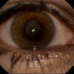
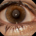
March 2002 – Eye exterior aspect
Looking back to a photo taken in March 2002, i can say that they eye regained a normal looking aspect after the vitrectomy abuse, it is indeed smaller, but not noticeably.
April 16, 2003 – Inspection
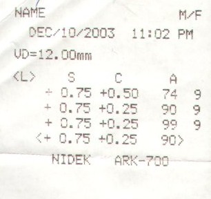
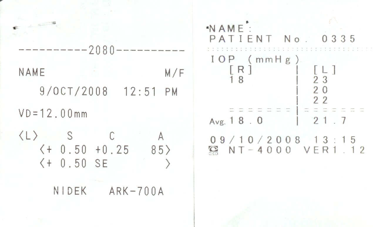
December 10, 2003 – Inspection
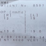
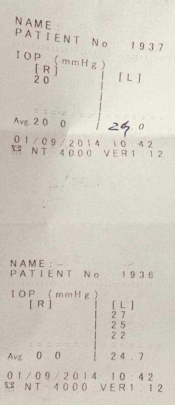
IntraOcular Pressure Test (IOP)
December 8, 2005
Treatment administrated:
- Maxidex, eye drops
- Gerovital Ochi, pills
Retrospecting the past
2002 – I remembered a scene like magic, which raised suspicion to me earlier, a few years ago (after 1997, but I can not say the exact year): one day, being in the kitchen, where the furniture is white, tiles on the floor are all white, as much light, I saw something moving with the eye for some time. At that time I did not insisted on observing the phenomenon, considering it was a mosquito or something. Was it back then all the effect of this affection/disease ?
October 2008 Status
- Right eye – posterior post-vitrectomy status: complicated cataract – surgical case exceeded
- Left eye – the only one functional, photocoagulated peripheral vasculitis
- peripheric lesions @ 10°° o’clock (2)
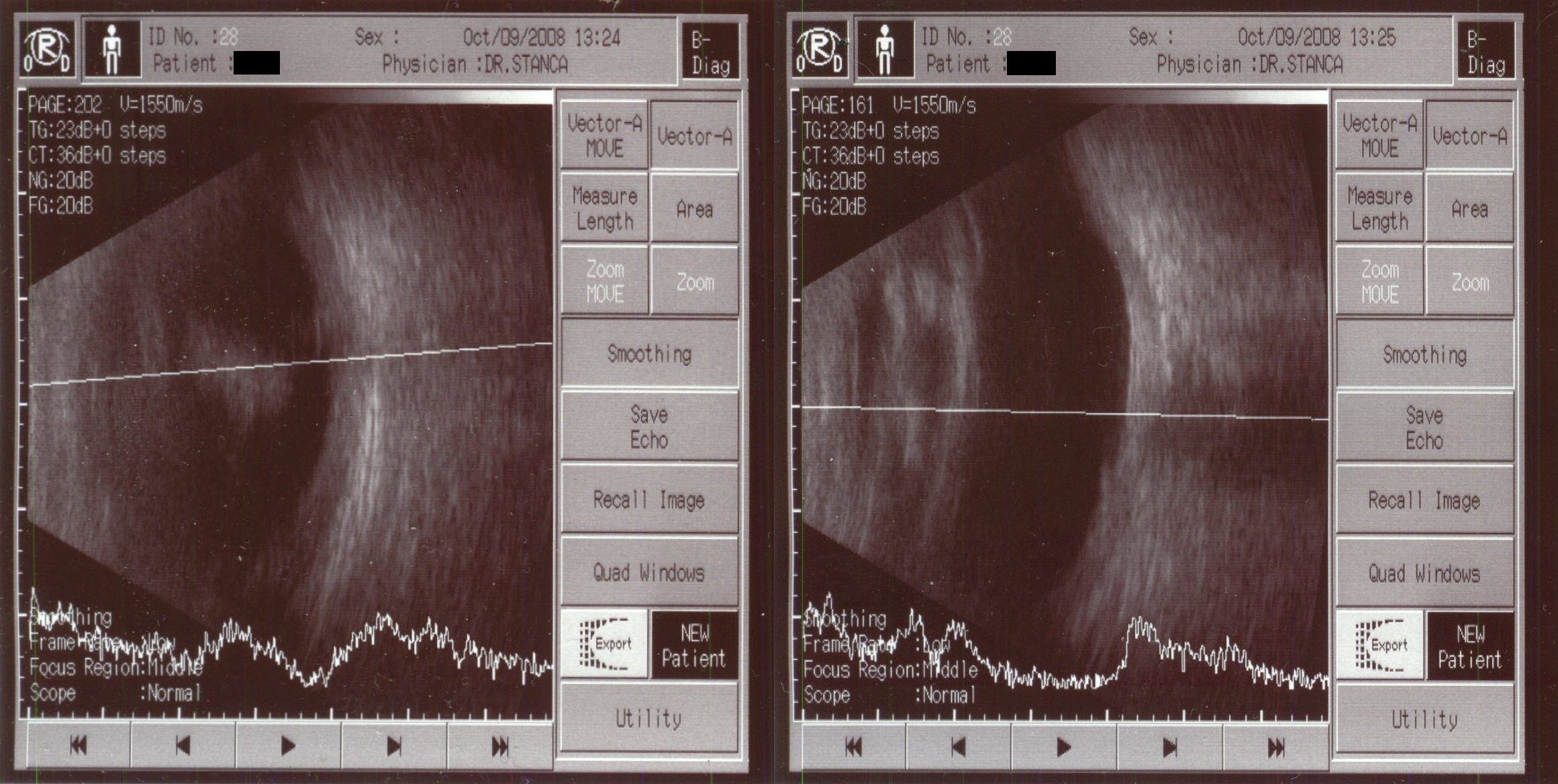
- Recommended periodical inspection at every 3 months, due to risks of vitreean hemoragy and retinal detachment in the left eye
- Treatment receipt received:
- Nutrof, 1cps/day, for 6 months
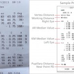
May 10, 2012
After stressing extended time working on computer, small dots in field of vision accentuated. I went to be consulted at WestEye Hospital. After consultation I was ensured that there is nothing wrong (?!) and all looks normal for the left eye.
Treatment received
- Redoxon 1000mg, 1tb/day, for 10 days
- Tarosin, 1tb three times/day, for 1 month
Following the consultation, I went for further investigation to another hospital where I received a light laser photocoagulation of some suspected areas (around nasal area.. as I was seeing the dots in the outer top right), from the same doctor.
March 2013 – Status
Other than symptoms described in the above representation, I am experiencing some retinal spasms, which manifest exactly the same as described by Scintillating scotoma, best matched by the following illustrations:


Symptoms of these spasm seems to manifest arbitrary to me, at an approx. interval of one to six months between (can’t identify a trigger), typically appear gradually over 5 to 20 minutes, expanding over half of the field of vision, with a random pattern and generally lasting fewer than 60 minutes, leading to a headache in the end, which lasts about an hour more max.
Having the right eye unfunctional, can’t tell if this may be a brain-superimposed image generated by the retina of other eye, or a Retinal Migraine. Will have to discuss this in detail with my doctor.
Watching this excellent video about Migraines: , cleared some of the fears:
- it is not a retinal detachment, as vision restores fully in fewer than 60 minutes
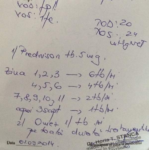
20 September 2013 – Inspection and Laser
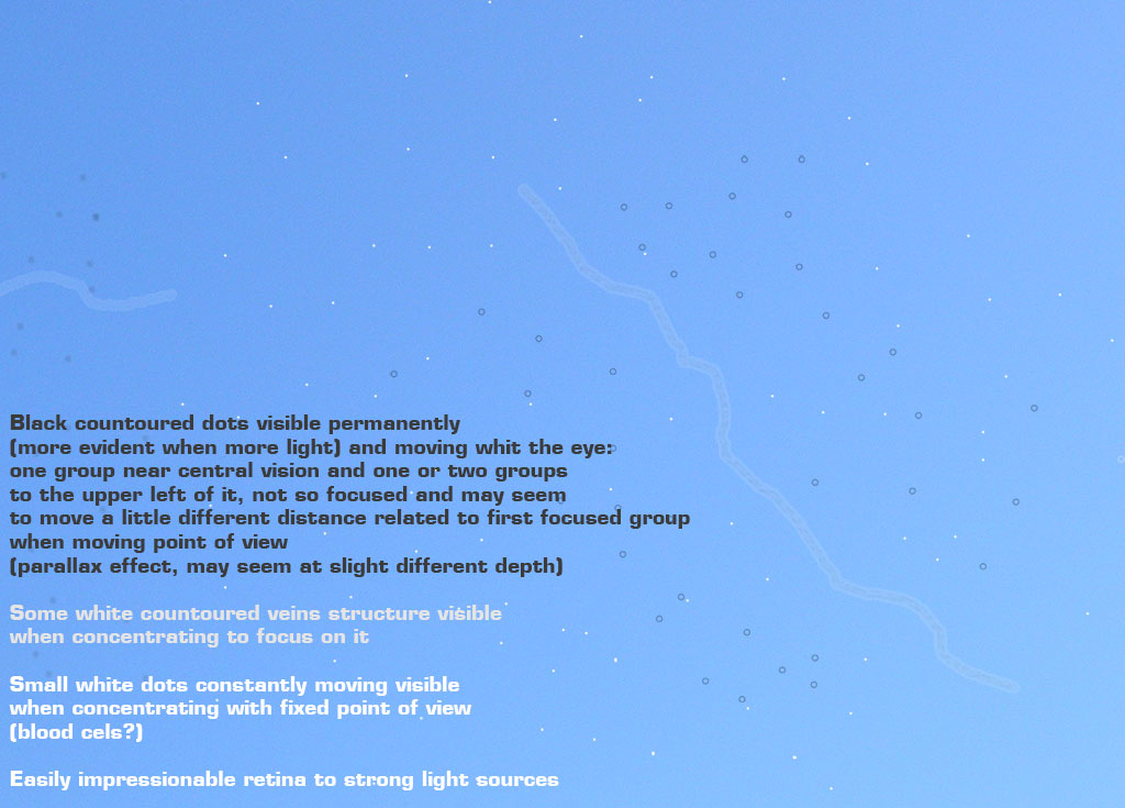
After one day and night of stress and no sleep on 1st of September, found the next day two big dots in the visual field as pictured:
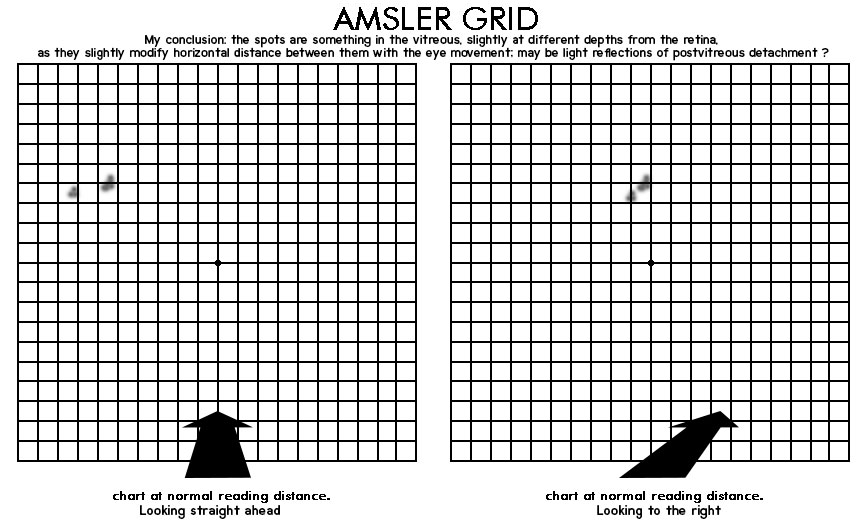
In the next 20 days monitored them against blue sky to see if they dissapear or changes appear. Found out that if I stare longer I can see a whole lot more very small dots spread all over the visual field, but only in strong light.
Medical inspection revealed a reactivated vasculitis in the periphery of the left eye, with an active retinal bleeding inside retina in the peripherical side with no risk of getting into the vitreous.
Laser was applied to isolate the area around, 816 dots at 12 o’clock position.
Treatment given:
- Medrol tb. 16mg, for 30 days, as follows:
- First 5 days: 3 tb. / day, all at once in the morning
- Day 6 to 10: 2 + 1/2 tb. / day, all at once in the morning
- Day 11 to 15: 2 tb. / day, all at once in the morning
- Day 16 to 20: 1 + 1/2 tb. / day, all at once in the morning
- Day 21 to 25: 1 tb. / day
- Day 26 to 30: 1/2 tb. / day
- Nexium, 1 tb./day for 30 days (as hepato protector)
- Mirtilene Ginko, 1 cps./day for 3 months
- Tarosin, 1 tb. x 3 / day for 3 months
Next inspection: 25 october 2013
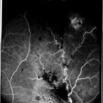
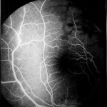
24 September 2013
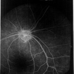
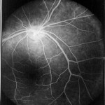
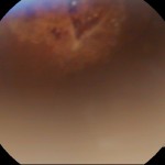
Eye Fundus Photos, does not show the current bleeding as the camera could not take images of the peripherical region
Left eye
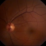

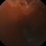
Right Eye
9 May 2014
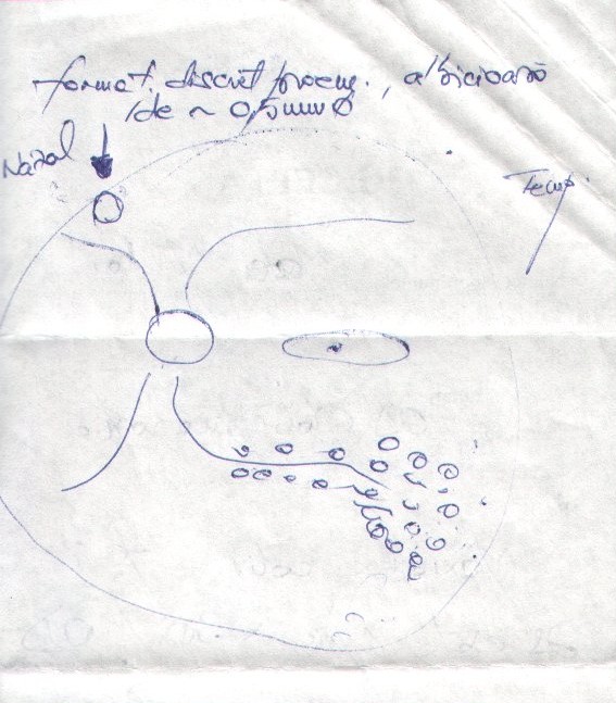
Eye fundus inspection
No pigment cells, no conglomerates, no retinal breakage
Inspection when eye looking leftmost: there are some fine vessel sheathing, beyond the area where I had laser last time. (In the temporal area, two white inflammation sprouts at the edge of temporal, easily sheathed vessels).
- Medrol tb. 16mg, for 30 days, as follows:
- First 3 days: 3 tb. / day, all at once in the morning
- Next 3 days: 2 + 1/2 tb. / day, all at once in the morning
- Next 3 days: 2 tb. / day, all at once in the morning
- Next 5 days: 1 + 1/2 tb. / day, all at once in the morning
- Next 5 days: 1 tb. / day
- Next 5 days: 1/2 tb. / day
- Omez , 1 tb./day for 30 days (as hepato protector)
- Mirtilene Ginko, 1 cps./day for 3 months
- Tarosin, 1 tb. x 3 / day for 3 months
Followup
A few laser points (6 / 8 / 10 points) should be made next time (on 6 June 2014), after inflammation will be gone. for the “muguri albiciosi pe prelungirea vasului”
No retinal bleeding now, as last time we had some..
6 June 2014 – After Medrol cure over
Vision almost clear, only a slight barely visible white contoured vessel.9
Eye fundus inspection
Revealed everything is fine and clean
- Mirtilene Ginko, 1 cps./day permanently, with 1 month break between 2 months
24 June 2014
Had two retinal migraines / scintillating scotoma in the last 2 days, around 11am-13pm (may be triggered by coffee)
Small dots reappear progressively in the visual field, along with contoured white vessels.
….
fast forward to more recent news, missing period to be filled soon and more details to be added
…
1 September 2014
Small dots indicated me that I should go inspecting the retina again. Seems there is still a small active vasculitis. Doctor says this will be the last session of corticosteroids, and this is given now just for my extra safety as there is no real risk from his point of view.
November 2014
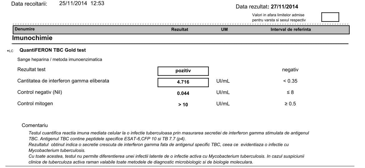
Following the study of prof. Dr. Jyotirmay Biswas, Director of Uveitis and Ocular Pathology departments – Sankara Nethralaya – Eales Disease – Current concepts in diagnosis and management,
I did a QuantiFERON TB Gold Test which came out positive!
It is unclear to me why at the stage of 2001, no one recommended a simple Tuberculosis analysis.
December 2015
I’ve started anti-tuberculous treatment, but excluding ethabuthol as my local doctor from Marius Nasta Institute considered for my case, of only one functional eye left.
February 2015
Switched to next stage of anti-TB treatment, with lower medication. 600mg isoniazide + rifampicine.
2 March 2015
Had a scintillating scotoma episode in the morning, after drinking some coffee (aura dissapeared in max 1 hour, headache at back of the head dissapeared after a few more hours with aid of Nurofen and resting)
14 March 2015
Woke up in the morning with pulsating light flashes coming from the nose area towards central vision. Tought it’s an scintillating scotoma and put myself immediately to rest, waiting for it to pass. Two days passed and no change. Permanent flashes all the weekend for 2 days.
16 March 2015
Got to ophtalmological consult.
BIG BREAKTHROUGH FINDING!
Retina looks better than ever in the last 15 years! It seems that the anti-TBC treatment was effective regarding the retina.
For the flashes, no cause could be found at eye inspection. Neurological consult was recommended.
Eye tension 22.
17? March 2015
Neurological consult recomments RMN (IRM) cerebral with details on optic nerves and contrast fluid.
Isoniazide may give optic nerve problems and this is the followed path for now.
21 March 2015
IRM result and interpretation indicate a fine deposit of contrast in the retrobulbar area of the optic nerve, on a length of about 5mm.
Anti-inflammatory treatment with Solu-Medrol (1gr for 3 days, 500mg for next 2 days) or oral Medrol is recommended rightaway.
Between 2015 – 2019 – To be updated..
No major concerns. In 2019, a Tinnitus showed up in both ears, cause cannot be tracked.
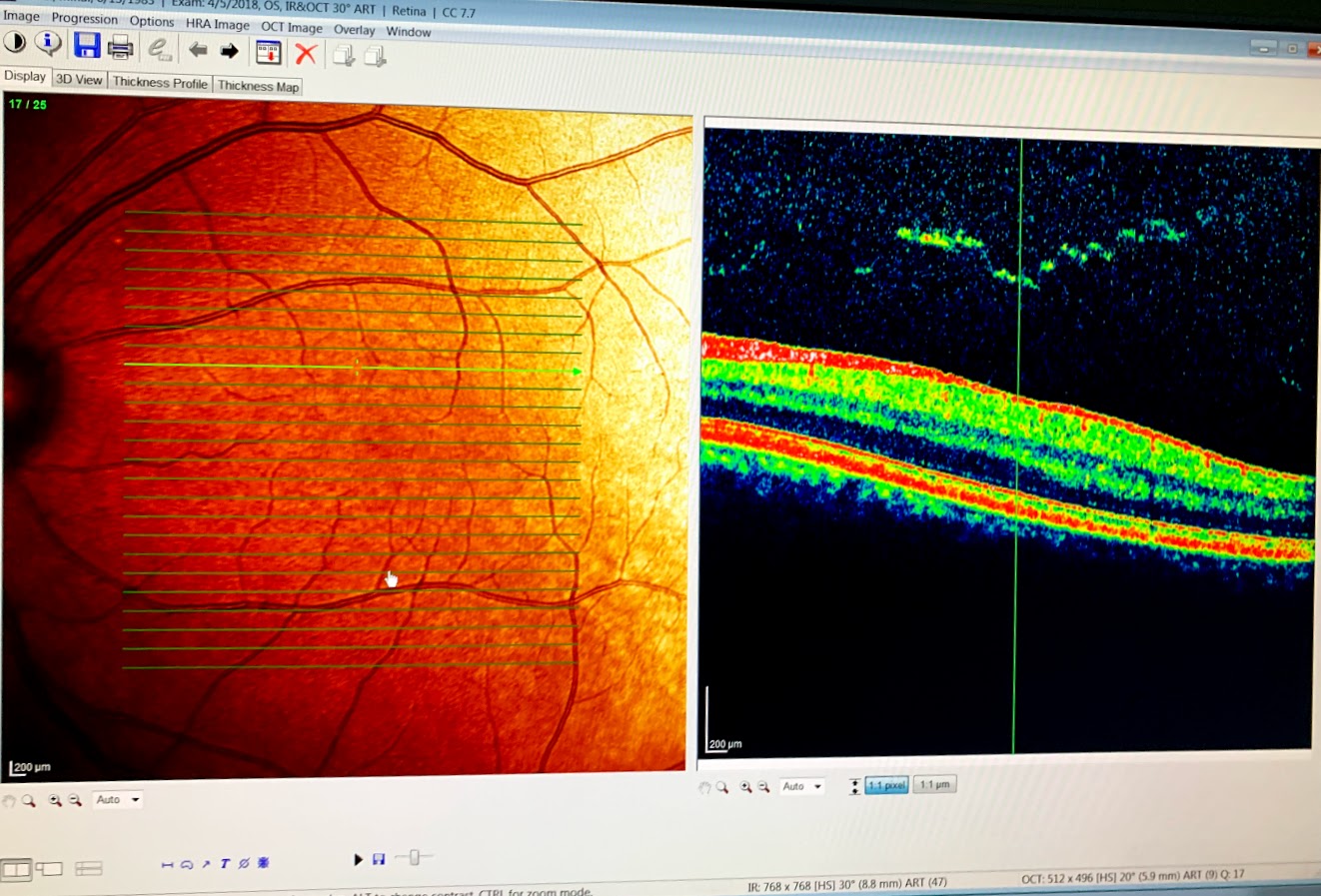
5 April 2018
OCT
Optical Nerve concerns
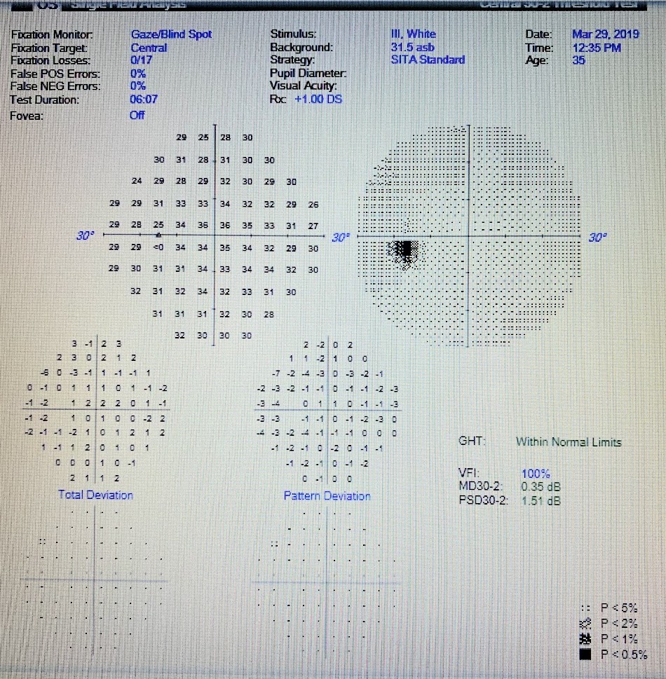
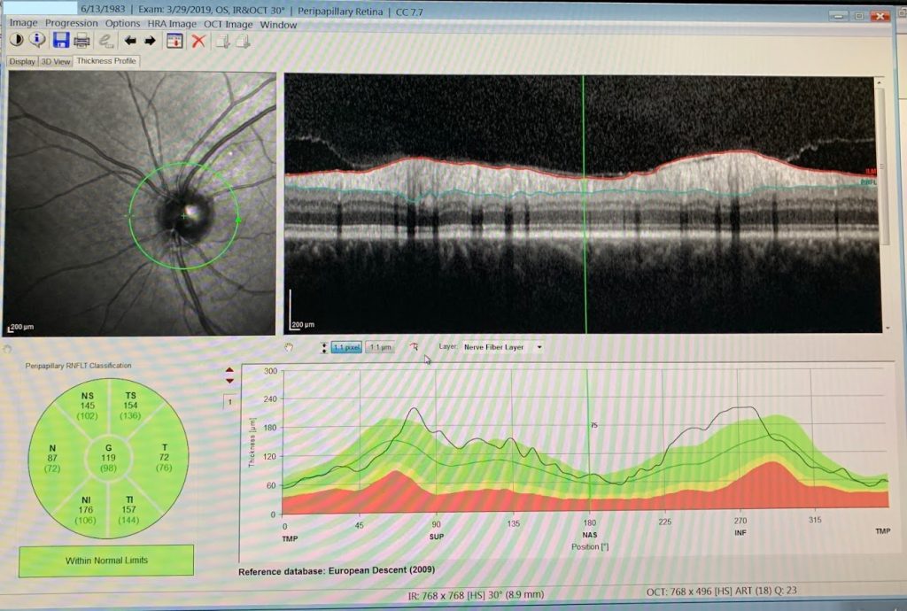
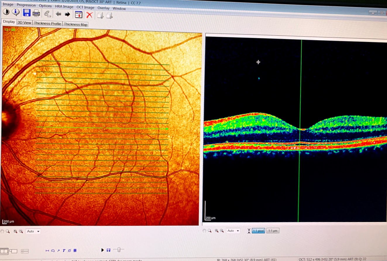
29 March 2019
FOV and OCT
21 May 2019
On 21 May 2019, after a lack of sleep night before a plane travel, combined with stress, next morning I’ve noticed upon wakeup, on a white wall, that there is a dark circle (black disk) appearing when transitioning from darkness to light (when blinking for example), which is fading away in a second. Also noticeable in the same position, it’s circular outline when moving eyes quick. Also felt vision in darkness decreased.
25 May 2019
Following dark circle symptoms from 21 May 2019,
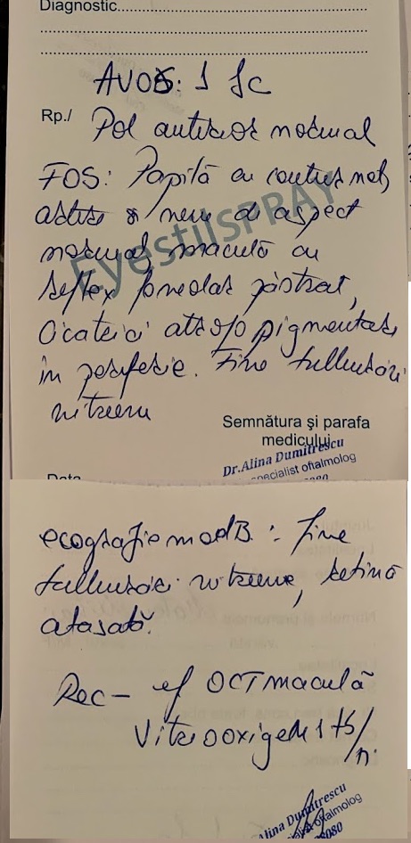
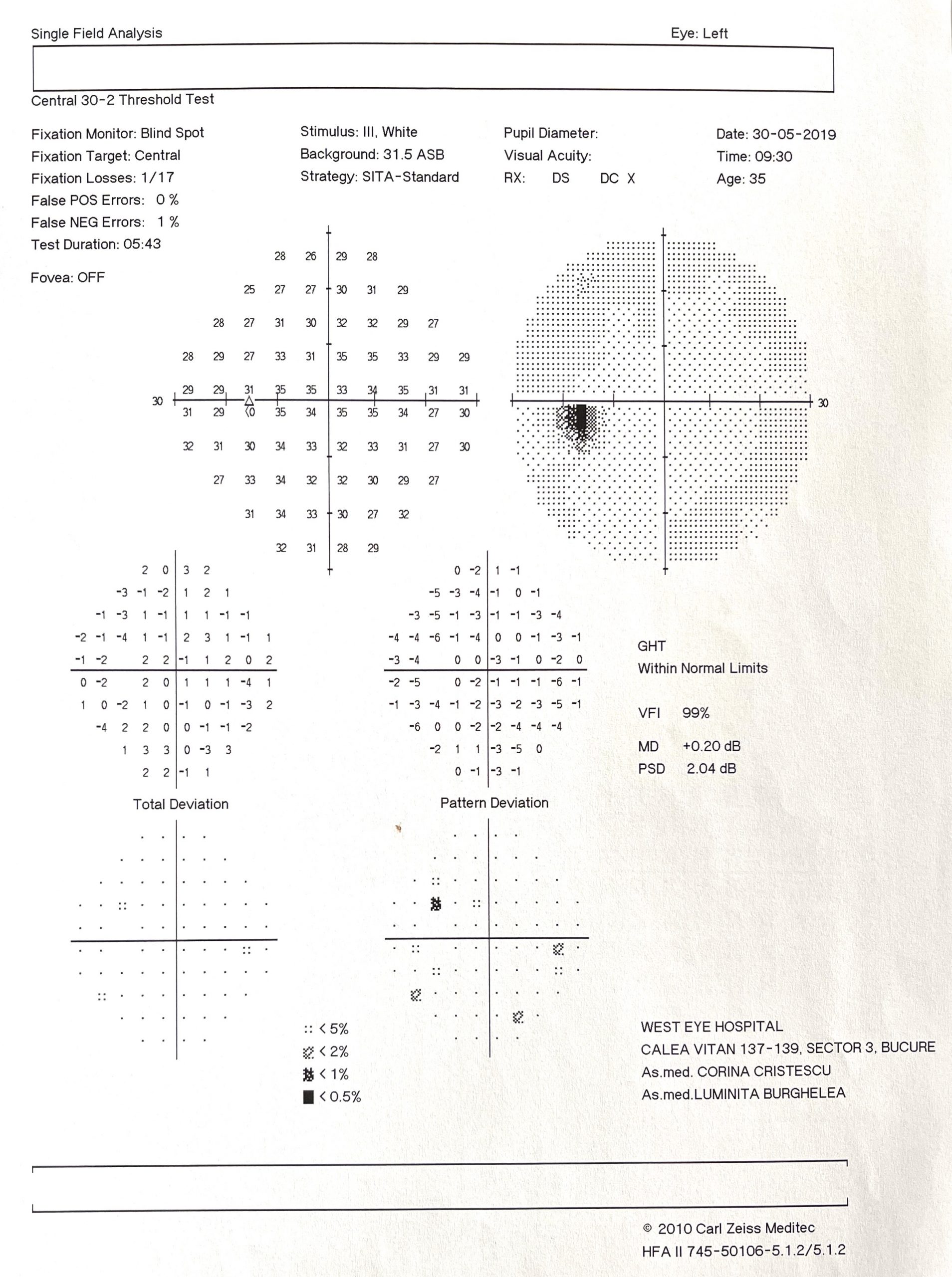
28 May 2019
Following dark circle symptoms from 21 May 2019,
FOV and OCT
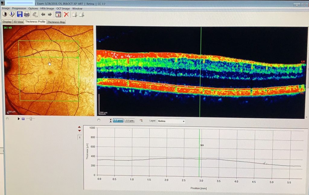
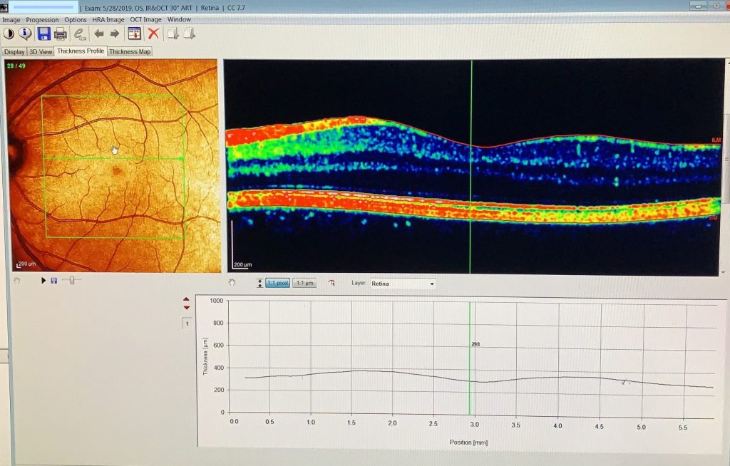
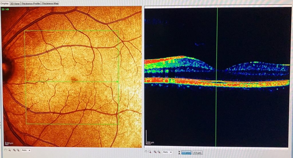
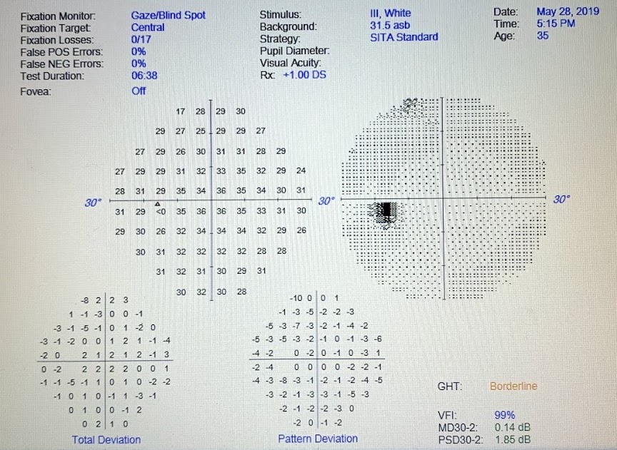
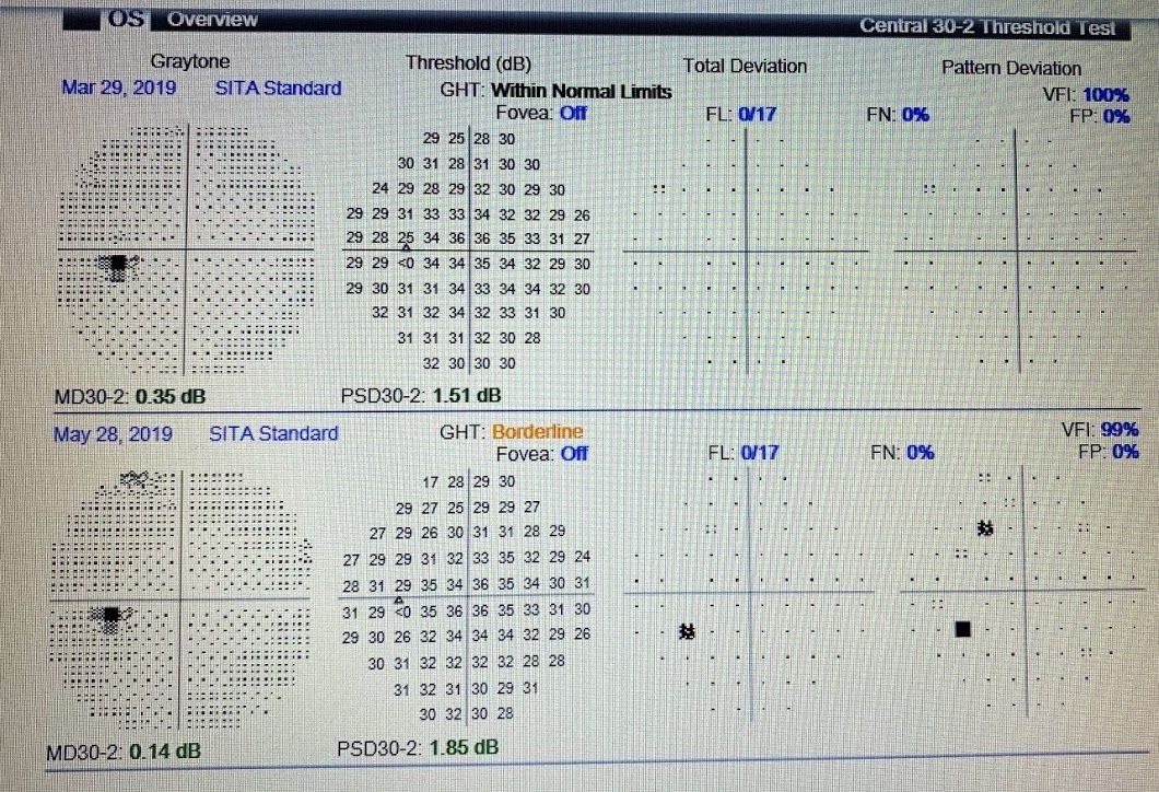
Notice the FOV compared to two months ago, in the last screenshot.
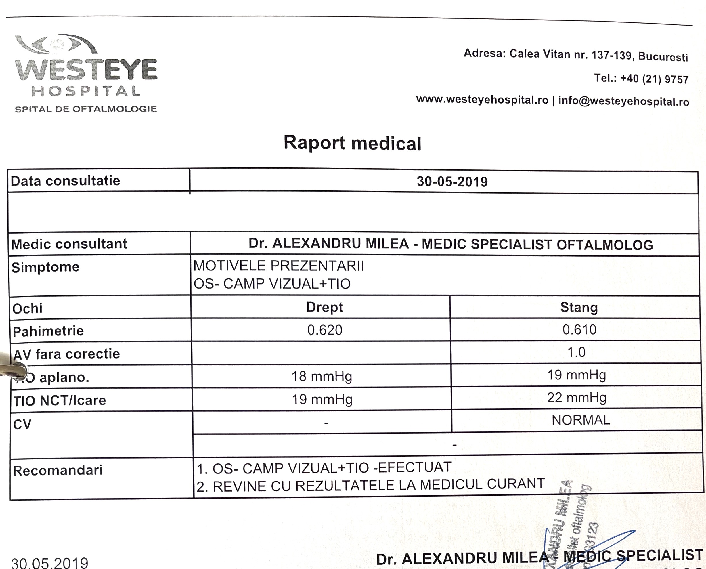
30 May 2019
FOV repeat and TIO
Followup – to be updated – dark circle symptom faded away in few months..
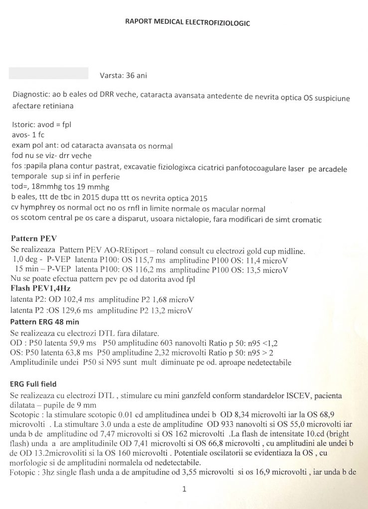
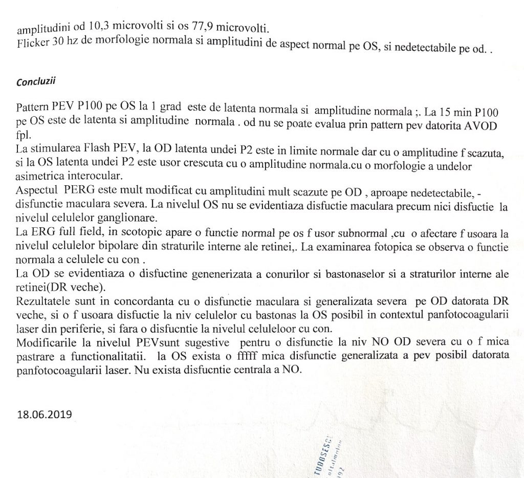
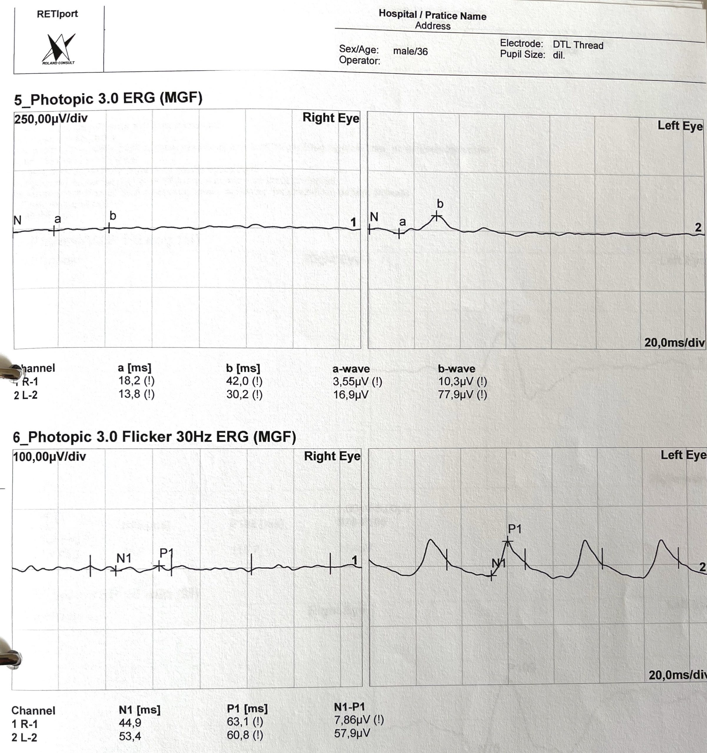
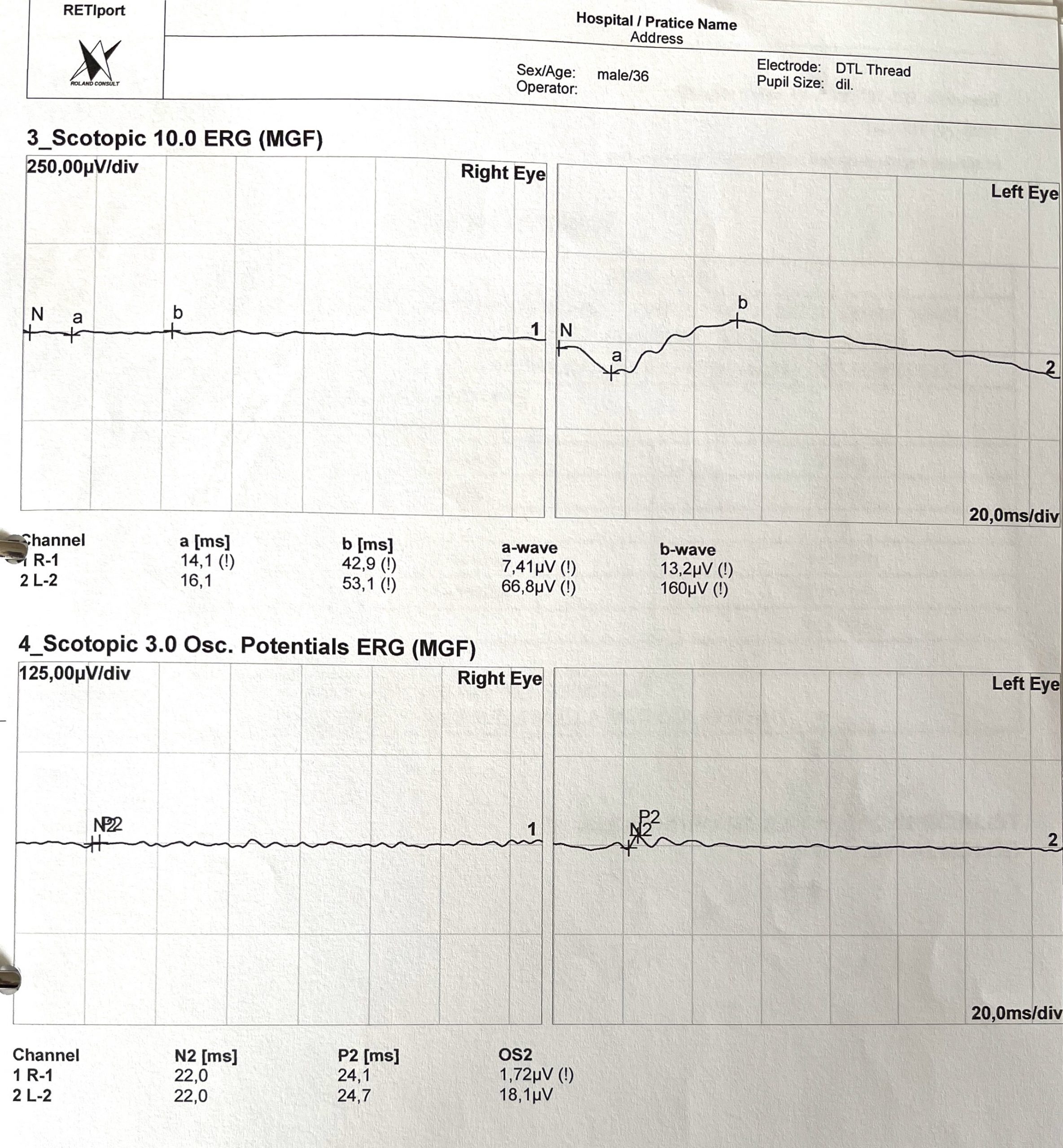
18 June 2019
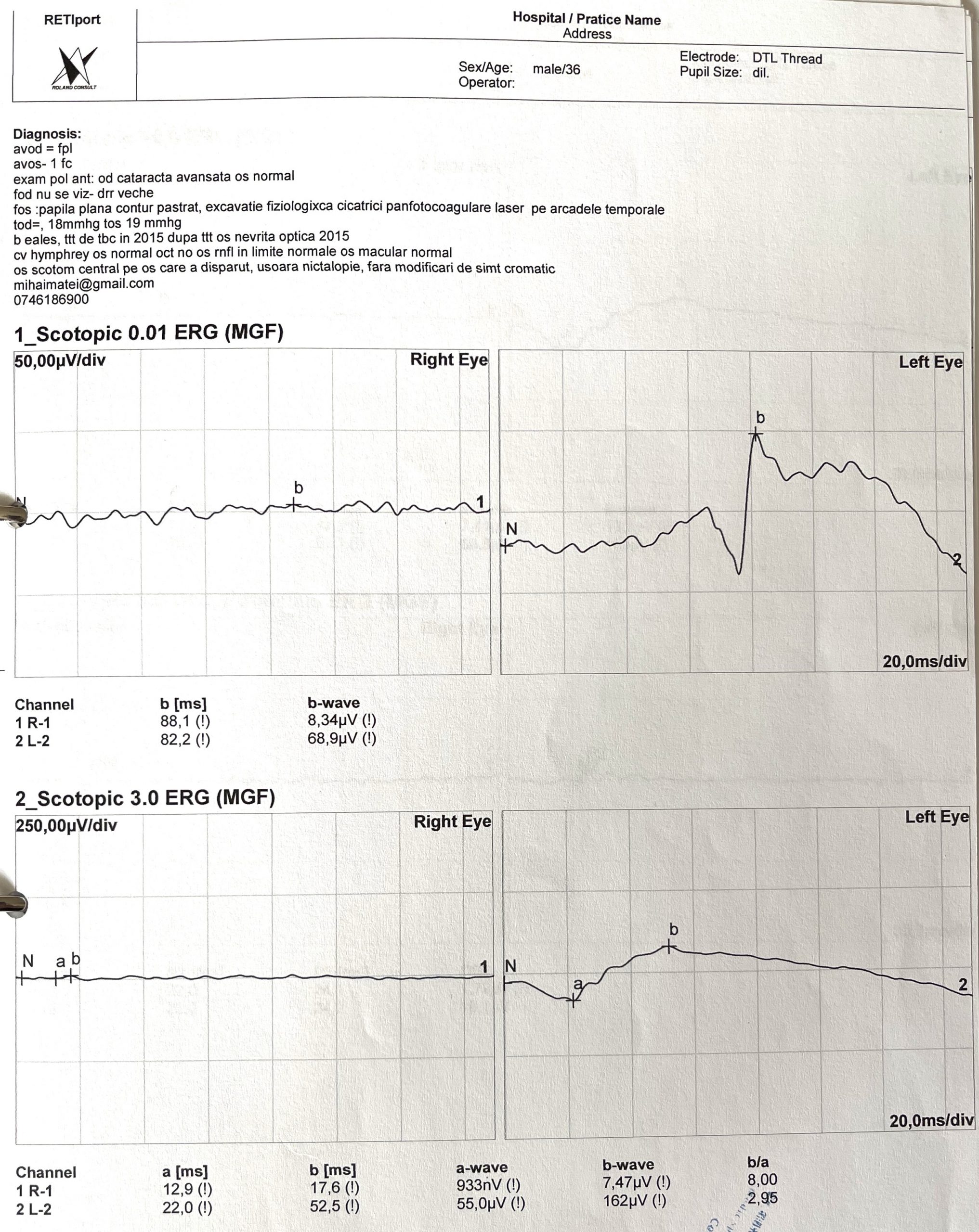
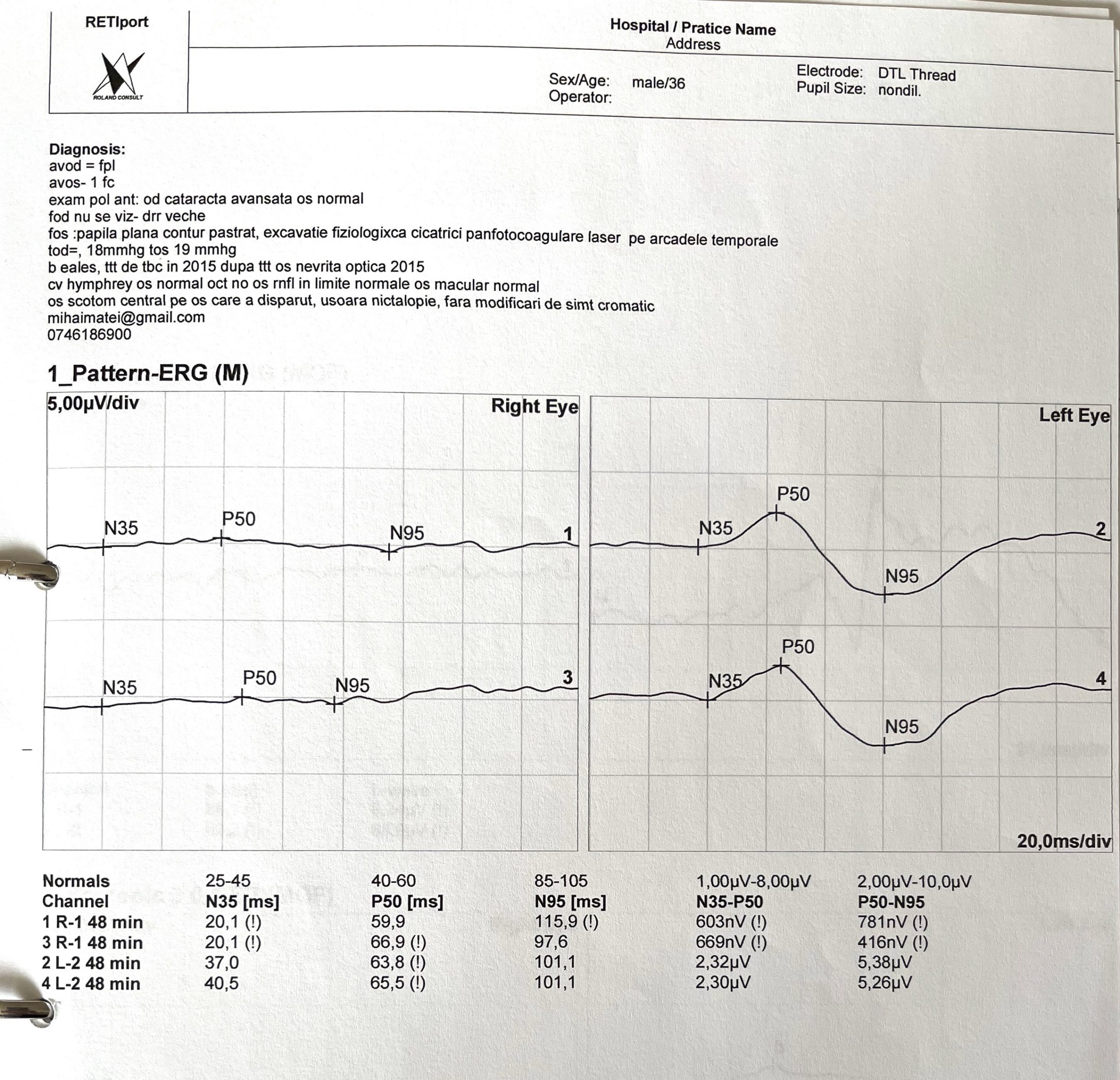
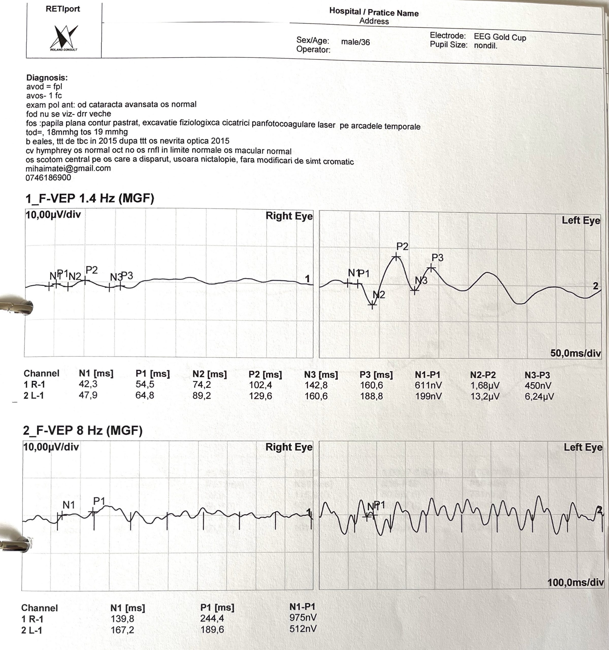
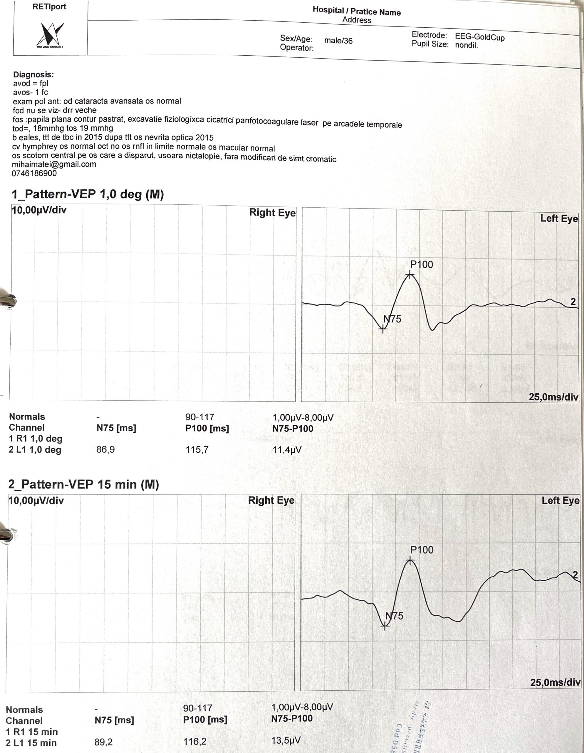
ERG – Electroretinogram
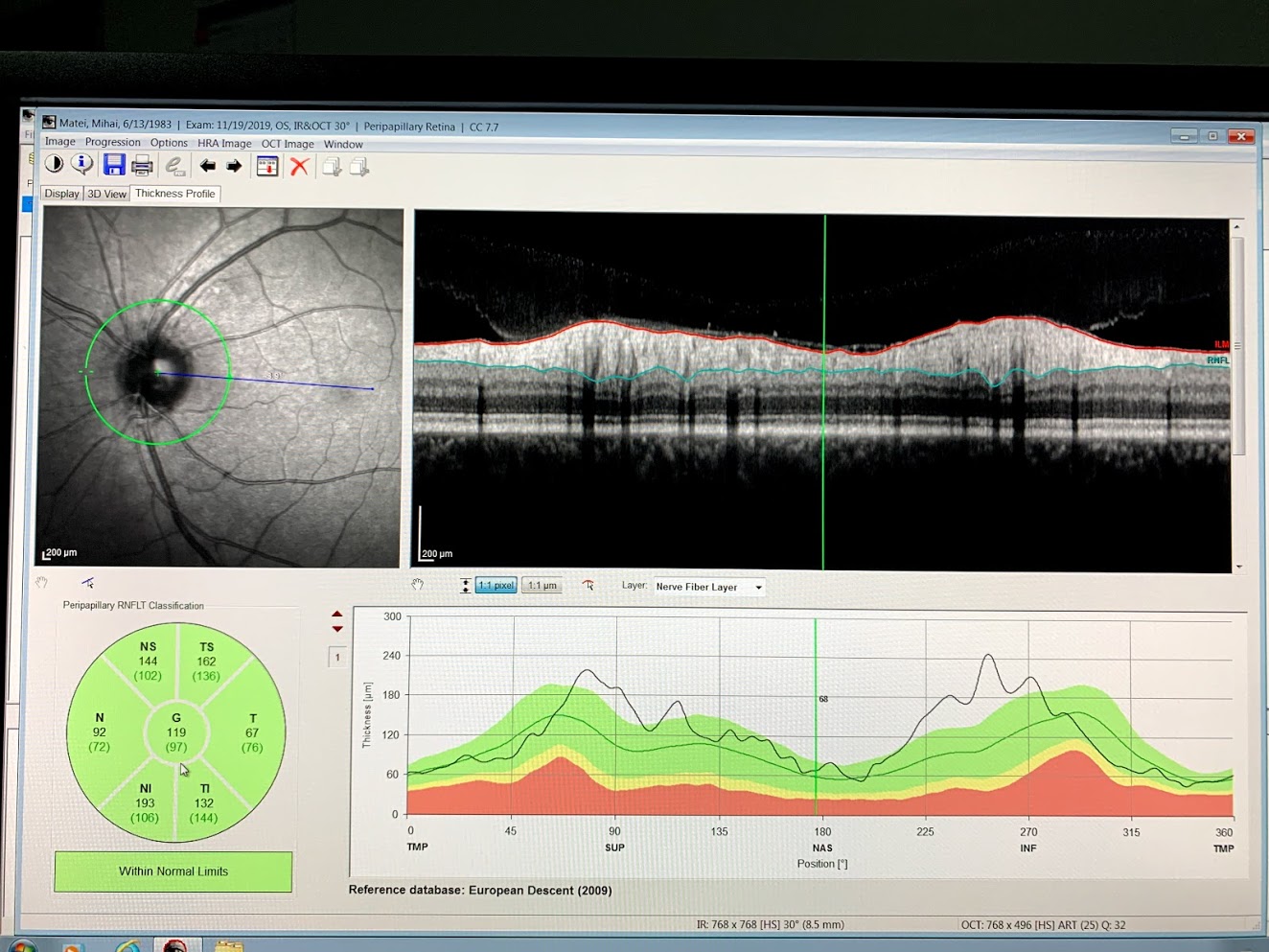
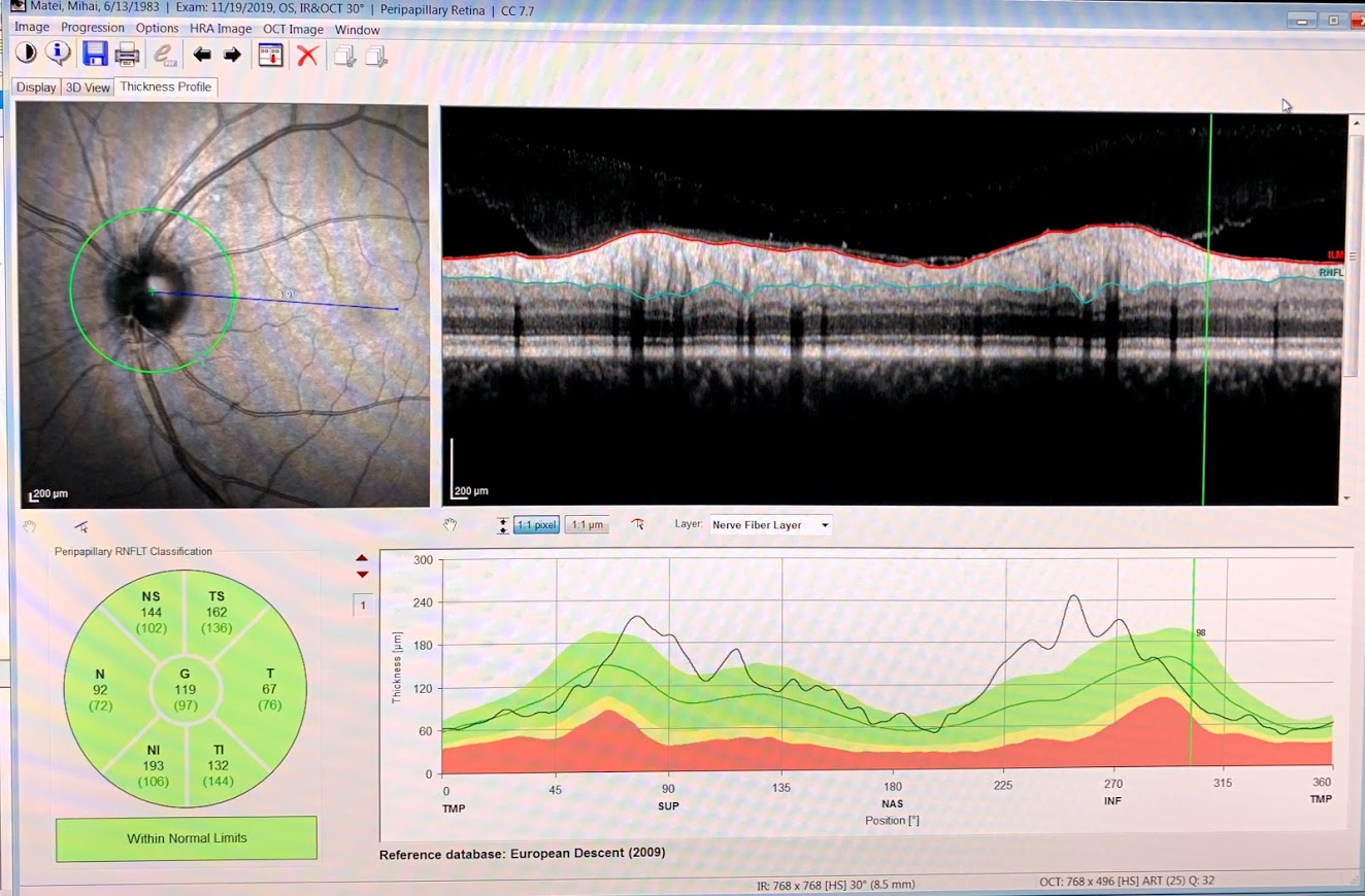
19 November 2019
OCT
28 January 2020
Regular checkup, no issues
31 March 2021
Again, on a lack of sleep/inconsistent sleeping hours and stress background, the dark circle showed up.
OCT and Eye Fundus Inspection
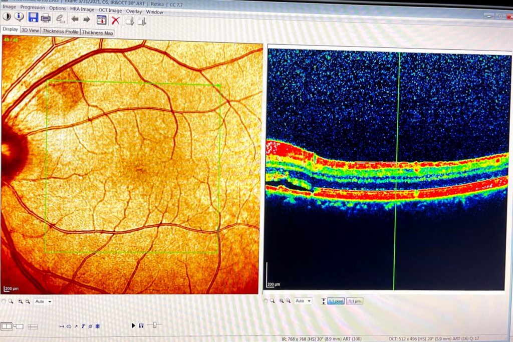
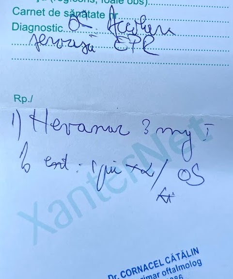
Eye Fundus Inspection does not reveal it.
Recommended treatment: Nevanac drops 2 times/day and another checkup after 2 weeks.
Link to a study related to Nevanac: https://www.ncbi.nlm.nih.gov/pmc/articles/PMC6699520/
This study links to another study, mentioning good results with low dosage Aspirin: Caccavale A, Imparato M, Romanazzi F, Negri A, Porta A, Ferentini F. A new strategy of treatment with low-dosage acetyl salicylic acid in patients affected by central serous chorioretinopathy. Med Hypotheses. 2009;73(3):435–437. doi:10.1016/j.mehy.2009.03.036